Details Of Human Eye Anatomy
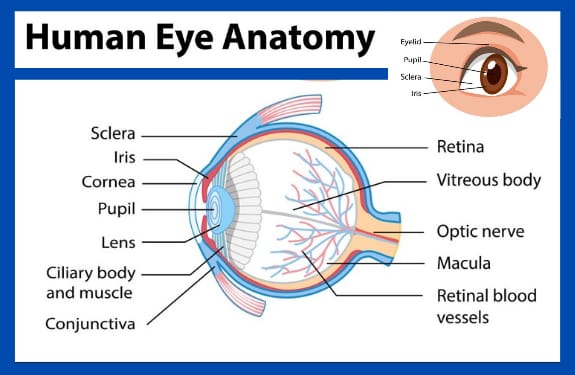
External Eye Structures:
1. Cornea: Transparent outer layer covering the eye
2. Sclera: White, tough outer layer protecting the eye
3. Iris: Colored part controlling light entry
4. Pupil: Opening in the iris regulating light
5. Eyelids: Upper and lower lids protecting the eye
6. Eyelashes: Hairs on the eyelids filtering debris
Internal Eye Structures:
1. Lens: Clear, flexible structure focusing light
2. Retina: Light-sensitive tissue converting light to signals
3. Macula: Central part of the retina for sharp vision
4. Optic Nerve: Carrying visual signals to the brain
5. Vitreous Humor: Clear gel filling the eye’s center
6. Aqueous Humor: Clear fluid nourishing the cornea and lens
Other Important Structures:
1. Conjunctiva: Membrane covering the white part of the eye
2. Choroid: Layer of blood vessels supplying the retina
3. Ciliary Muscles: Muscles controlling lens shape
4. Fovea: Small pit in the macula for central vision
Key Points:
1. The eye is a complex, sensitive organ.
2. Each structure plays a vital role in vision.
3. Understanding eye anatomy helps appreciate vision and eye health.
Cornea: Transparent outer layer covering the eye
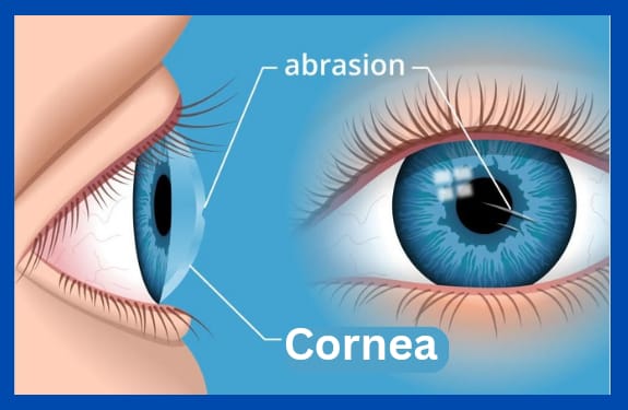
The cornea is the transparent, dome-shaped surface at the front of the eye. It is the outermost layer of the eye and plays a crucial role in vision. The cornea:
1. Refracts (bends) light entering the eye
2. Provides most of the eye’s focusing power
3. Protects the eye from dust, dirt, and other foreign particles
The cornea is made up of three layers:
1. Epithelium (outer layer)
2. Stroma (middle layer)
3. Endothelium (inner layer)
The cornea is:
1. Transparent and avascular (no blood vessels)
2. Richly innervated (supplied with nerves) for sensitivity
3. Self-healing and regenerative
The cornea is essential for clear vision, and any damage or disease affecting the cornea can impact vision and eye health.
Functions of the cornea:
1. Refraction
2. Protection
3. Sensation
4. Lubrication
5. Clarity
6. Focusing power
7. Eye pressure regulation
8. Aiding tear distribution
9. Supporting eye movements
10. Maintaining eye shape
Overall, the cornea is a vital part of the eye, and its health is essential for maintaining good vision and eye comfort.
Sclera: White, tough outer layer protecting the eye
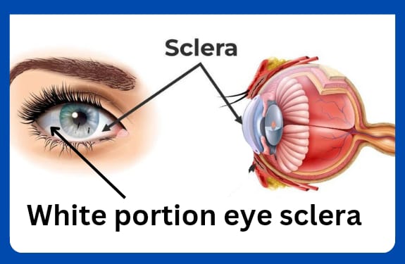
The sclera is the white, tough, outer layer of the eye that provides protection and structure to the eye. Here are some key facts about the sclera:
1. Location: The sclera covers about 80% of the eye’s surface, starting from the cornea (the transparent front layer) and extending to the optic nerve (which carries visual information to the brain).
2. Function: The sclera provides:
– Protection: shields the eye from injury and infection
– Structure: maintains the eye’s shape and size
– Attachment: serves as an attachment point for eye muscles
3. Composition: The sclera is made of:
– Collagen fibers (tough, fibrous proteins)
– Elastic fibers (allowing for flexibility)
– Ground substance (a gel-like material)
4. Thickness: The sclera is approximately 0.3-0.4 mm thick in the anterior (front) region and 0.5-0.6 mm thick in the posterior (back) region.
5. Color: The sclera appears white due to the scattering of light by the collagen fibers.
6. Blood supply: The sclera has a limited blood supply, which helps to maintain the eye’s clarity.
7. Nerve supply: The sclera has nerve endings that detect pain, pressure, and touch.
8. Diseases: Scleral disorders include:
– Scleritis (inflammation)
– Episcleritis (inflammation of the outer layer)
– Scleral thinning or perforation (due to injury or disease)
The sclera plays a vital role in maintaining the eye’s integrity and function.
Iris: Colored part controlling light entry
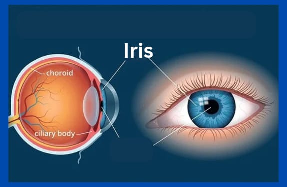
The iris is the colored part of the eye that surrounds the pupil, controlling the amount of light that enters the eye. Here are some interesting facts about the iris:
1. Color: The iris is responsible for eye color, which is determined by the amount and distribution of pigment.
2. Structure: The iris is made up of two layers:
– Stroma (front layer)
– Epithelium (back layer)
3. Function: The iris regulates light entry by adjusting the pupil size.
4. Muscles: The iris contains two types of muscles:
– Dilator pupillae (enlarges pupil)
– Sphincter pupillae (constricts pupil)
5. Pupil control: The iris controls pupil size, influencing:
– Light entry
– Depth of field
– Focus
6. Unique patterns: Iris patterns are unique to each individual, like fingerprints.
7. Diseases: Iris disorders include:
– Iritis (inflammation)
– Iris melanoma (cancer)
– Anisocoria (unequal pupil sizes)
8. Surgery: Iris surgery can correct conditions like iris defects or tumors.
9. Symbolism: The iris is often associated with:
– Greek mythology (Iris, the messenger goddess)
– Art and literature (symbolizing beauty, hope, and wisdom)
The iris plays a vital role in vision and eye health, making it a fascinating and important part of the human eye!
Pupil: Opening in the iris regulating light
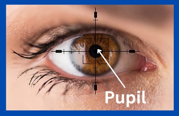
The pupil is the opening in the center of the iris that regulates the amount of light entering the eye. Here are some interesting facts about the pupil:
1. Size control: The pupil changes size to control light entry, dilating (enlarging) in low light and constricting (reducing) in bright light.
2. Muscle control: The pupil is controlled by two muscles:
– Sphincter pupillae (constricts pupil)
– Dilator pupillae (dilates pupil)
3. Light reflex: The pupil responds to light through the pupillary light reflex, constricting when light enters the eye.
4. Shape: The pupil is typically round, but can appear irregular in certain conditions.
5. Size variation: Pupil size varies among individuals, with an average diameter of 2-4 mm.
6. Adaptation: The pupil adapts to changes in light levels, taking about 5 minutes to fully adjust.
7. Emotional response: Pupil dilation can be an indicator of emotional arousal, such as excitement or fear.
8. Neurological indicator: Abnormal pupil responses can indicate neurological disorders, such as coma or brain injury.
9. Age-related changes: Pupil size decreases with age, affecting low-light vision.
10. Unique feature: Like fingerprints, no two people have the same pupil shape or size!
The pupil plays a vital role in regulating light entry and is an important aspect of eye health and function.
Eyelids: Upper and lower lids protecting the eye
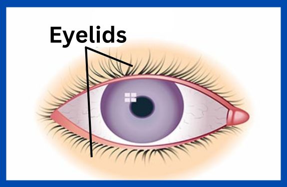
The eyelid, also known as the palpebra, is a thin, movable fold of skin that covers and protects the eye. Here are some interesting facts about the eyelid:
1. Function: Eyelids:
– Protect the eye from dust, debris, and injury
– Regulate light entry
– Help distribute tears
– Aid in eye movement
2. Structure: The eyelid consists of:
– Skin
– Muscles (orbicularis oculi and levator palpebrae)
– Connective tissue
– Tarsal plate (a thin, cartilaginous plate)
– Meibomian glands (oil-producing glands)
3. Types: There are two eyelids:
– Upper eyelid (larger and more mobile)
– Lower eyelid (smaller and less mobile)
4. Movements: Eyelids can move:
– Upward (elevation)
– Downward (depression)
– Laterally (abduction)
– Medially (adduction)
5. Blinking: Eyelids blink:
– Approximately 12-15 times per minute
– To distribute tears and lubricate the eye
– To protect the eye from irritants
6. Conditions: Eyelid disorders include:
– Blepharitis (inflammation)
– Entropion (eyelid rolls inward)
– Ectropion (eyelid rolls outward)
– Ptosis (droopy eyelid)
7. Surgery: Eyelid surgery can correct:
– Cosmetic issues (blepharoplasty)
– Functional problems (ptosis repair)
8. Age-related changes: Eyelids can experience:
– Sagging
– Thinning
– Loss of fat and muscle tone
The eyelid plays a vital role in protecting and maintaining eye health, and its movements are essential for clear vision and comfort.
Eyelashes: Hairs on the eyelids filtering debris
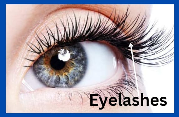
Eyelashes are the hairs that grow along the edge of the eyelid, protecting the eye from dust, debris, and other foreign particles. Here are some interesting facts about eyelashes:
1. Function: Eyelashes:
– Act as a barrier against dust, dirt, and other irritants
– Help to filter out light
– Aid in detecting objects close to the eye
– Play a role in eye health and hygiene
2. Structure: Eyelashes are made of:
– Keratin (a protein)
– Follicles (rooted in the eyelid)
– Shaft (visible part)
– Root (hidden part)
3. Growth: Eyelashes:
– Grow in cycles (anagen, catagen, telogen phases)
– Take about 3-4 months to grow
– Can grow up to 1-2 cm (0.4-0.8 in) long
4. Number: Most people have:
– Upper eyelid: 100-150 lashes
– Lower eyelid: 50-75 lashes
5. Types: There are two types of eyelashes:
– Cilia (short, fine lashes)
– Guard hairs (longer, thicker lashes)
6. Care: To maintain healthy eyelashes:
– Gently clean the eyelids
– Avoid excessive rubbing or pulling
– Use mascara and makeup remover carefully
7. Loss: Eyelash loss can be caused by:
– Aging
– Medical conditions (e.g., alopecia, blepharitis)
– Injury or trauma
– Poor eye care
8. Enhancement: Eyelashes can be enhanced with:
– Mascara
– False lashes
– Lash extensions
– Lash growth serums
Eyelashes play a vital role in protecting and beautifying our eyes!

Very nice for share knowledge